Lead Diaphragms
Jaytee Alloys: Lead Diaphragm
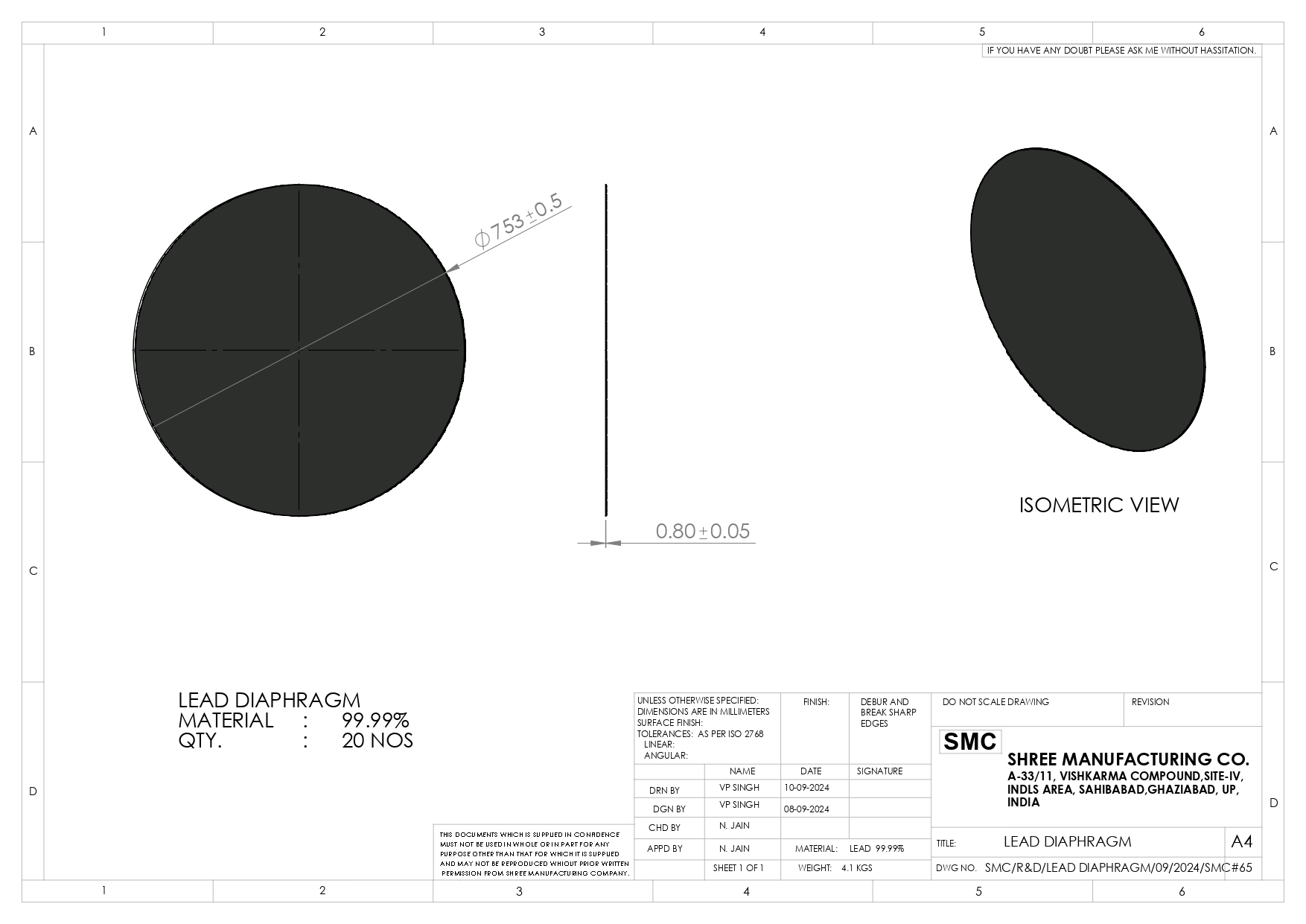
A lead diaphragm is a collimating device used in X-ray imaging to restrict the X-ray beam, reducing scattered radiation and protecting sensitive tissues. It works by using lead, or lead-rubber materials, to create specific, often rectangular, fields for imaging. This technology improves image contrast and safety.
Key Uses of Lead Diaphragms in X-ray Imaging:
-
Beam Collimation: Limits the size of the X-ray beam to only the area of interest to reduce the patient dose.
-
Scatter Reduction: Minimizes scattered radiation during radiographic projections, such as lateral elbow exams, by adding extra shielding to standard collimators.
-
Dose Reduction: Helps protect radiosensitive organs by reducing the amount of scatter from the main beam, note.
-
Customization: They are used in conjunction with "master cones" or light beam diaphragms (LBDs) in radiographic equipment, explainand this ScienceDirect article.
Important Considerations:
-
Positioning: The efficiency of a lead diaphragm varies depending on its distance from the X-ray source; it is generally more effective when placed farther from the source.
-
Beam Quality: While reducing scatter, they must be properly designed to avoid unintended beam shaping or irregular field shapes.


Our Certifications